Vadzo Imaging announces the Falcon-821CRS, a compact 8MP 4K microscope USB camera engineered for digital pathology, brightfield microscopy, and fluorescence imaging. Centered on the Onsemi AR0821 Back-Side Illuminated CMOS sensor with Linear HDR architecture, the module delivers consistent color accuracy and high dynamic range output across stained tissue histology, immunohistochemistry, and cytology slide imaging workflows through a USB 3.2 interface with UVC compliance.
FORT WORTH, TX / ACCESS Newswire / July 13, 2026 / Vadzo Imaging, a developer of embedded vision camera modules for medical, laboratory, and scientific imaging applications, has announced the availability of the Falcon-821CRS as an AR0821 Microscope Camera suited for brightfield pathology and fluorescence microscopy environments. The module combines the Onsemi AR0821 Back-Side Illuminated CMOS sensor with a USB 3.2 UVC interface, delivering 4K HDR imaging capabilities for digital pathology, histology, and cytology workflows across Windows, Linux, and Android host platforms without driver installation requirements.
Imaging Challenges in Pathology and Laboratory Microscopy
Pathology laboratories and research microscopy environments impose illumination conditions that differ substantially between modalities. Transmitted brightfield slide examination operates at a stable high intensity light level, while fluorescence excitation produces emission signals at the sensor plane that fall several orders of magnitude below brightfield intensity levels. A camera module operating as a Slide Illumination USB Camera must accommodate this intensity variation without per-frame manual reconfiguration. Platforms that require separate exposure calibration profiles for brightfield and fluorescence acquisition cannot function as a Consistent Illumination Camera across both modalities within the same imaging session, introducing inconsistency that complicates pathologist review and automated image analysis workflows that depend on normalized illumination baselines.
Stained biological specimens introduce a separate class of requirements centered on spectral accuracy. Hematoxylin and eosin preparations depend on contrast between blue violet nuclear staining and pink cytoplasmic regions, and a Stained Tissue USB Camera used in histology review must reproduce these spectral contrasts with sufficient channel linearity to support both visual and algorithmic assessment. Immunohistochemistry preparations introduce additional chromogenic markers whose separation depends on accurate color channel balance across the blue and red spectral bands, and an IHC Imaging Camera applied to DAB and Fast Red chromogen detection must maintain spectral fidelity without aggressive automatic white balance correction that distorts the ratio between stain channels and tissue background. A Histopathology Camera Module must also meet the lens interface geometry constraints imposed by laboratory microscope trinocular outputs, adding mechanical integration requirements beyond sensor performance criteria alone.
Subcellular morphological assessment requires spatial resolution beyond what consumer or standard industrial sensors deliver at typical microscope magnification levels. Nuclear membrane integrity, chromatin texture, mitotic figure identification, and cellular border irregularity are evaluated at 40x and higher magnifications, where pixel density determines diagnostic confidence. A Pathology Slide Camera configured for routine biopsy review must deliver sufficient resolution per field of view to resolve these features without tile reconstruction overheads that reduce acquisition throughput in high-volume clinical settings. For Tissue Imaging Camera applications where large numbers of slides must be processed per session, the sensor megapixel count and frame rate specifications directly affect system throughput and, therefore, the clinical utility of the imaging platform.
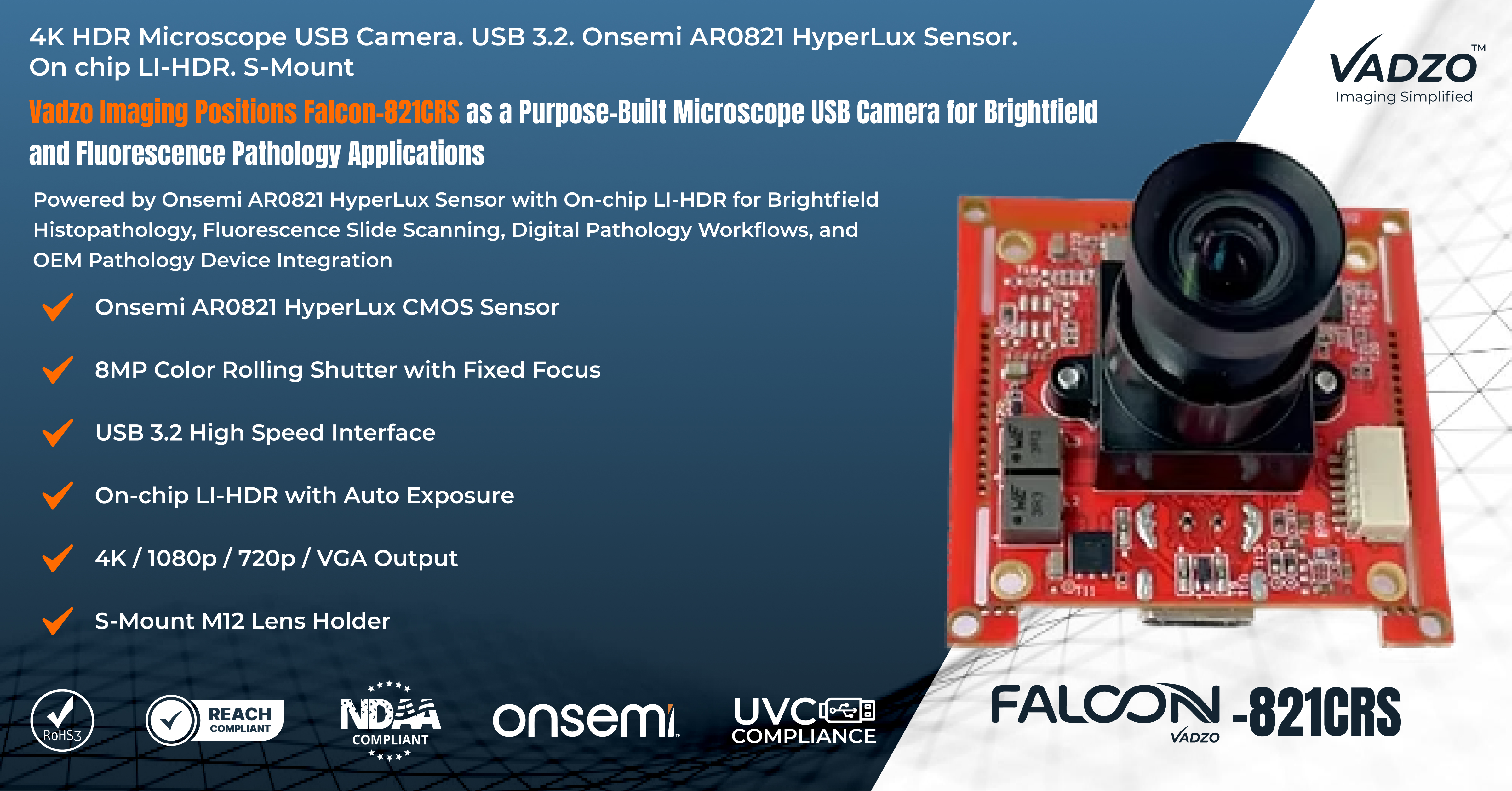
Falcon-821CRS: Compact Microscope USB Camera Module for Pathology and Fluorescence Imaging
The Falcon-821CRS is a compact M12 mount Microscope USB Camera module developed by Vadzo Imaging to serve brightfield pathology, fluorescence microscopy, and digital slide analysis applications within a single hardware configuration. The AR0821 Pathology Camera design centers on the Onsemi AR0821 Pathology Camera sensor - a Backside Illuminated CMOS device with an 8MP (3848 x 2168) array, 2.1 µm pixel pitch, and 1/1.7-inch optical format. Key specifications: Sensor: Onsemi AR0821 BSI CMOS | Resolution: 8MP (3848 x 2168) | Interface: USB 3.2 UVC | Lens Mount: M12 S-Mount | Dynamic Range: >90 dB (LI-HDR) | Platform: Windows, Linux, Android. As a 4K Microscope USB Camera with Linear HDR dynamic range exceeding 90 dB, the module handles the full illumination range of clinical brightfield and fluorescence imaging without per-frame exposure adjustment. Vadzo Imaging supplies the 8MP Pathology USB Camera as an OEM component ready for integration into clinical imaging instruments, laboratory workstations, and slide scanning platforms. The module operates as a Digital Microscopy Camera within compact instrument assemblies and as a 4K Pathology Camera reference platform for medical imaging product development, with M12 S-Mount lens interface accepting standard C-mount microscope relay adapters for direct coupling to trinocular microscope outputs.
Engineering Capabilities and Design Advantages
LI-HDR Architecture for Brightfield and Fluorescence Microscopy: Fluorescence and brightfield illumination regimes present intensity conditions that can differ by several orders of magnitude depending on the excitation source, emission filter, and objective in use. A AR0821 Brightfield Camera configuration without on-chip HDR processing cannot accommodate both regimes within a single exposure setting, requiring manual reconfiguration between brightfield and fluorescence acquisition cycles that reduces workflow throughput. The Falcon-821CRS operates as an HDR Microscopy Camera through the AR0821 sensor's Linear HDR (LI-HDR) readout architecture, which captures two exposure levels within the same on-chip readout cycle and produces a composited output frame without motion artifacts. This approach makes the Falcon-821CRS effective as a Brightfield Camera Module for high-intensity transmitted light imaging and simultaneously as a Fluorescence Camera Module for low-emission fluorophore detection, without hardware or firmware reconfiguration between modes. The LI-HDR output preserves highlight detail in high-intensity brightfield background regions while retaining signal information in weakly fluorescent emission channels within the same captured frame.
Back-Side Illuminated Pixel Architecture for Low Light Specimen Imaging: Fluorescence microscopy produces low photon flux at the sensor plane, particularly in configurations with low numerical aperture objectives, narrow bandpass emission filters, or specimens with low fluorophore labeling density. Conventional image sensor pixel architectures where metal routing layers occupy the path between the optical surface and the photodiode reduce quantum efficiency at shorter visible wavelengths by absorbing or scattering incident photons before they reach the photodiode active region. The Onsemi AR0821 employs a Backside Illuminated pixel architecture that removes routing structures from the photon collection path, improving quantum efficiency across the visible spectral range and reducing effective read noise at equivalent exposure durations. This architecture makes the Falcon-821CRS effective as a Fluorescence Microscopy Camera for wide field applications, including immunofluorescence labeling, DAPI nuclear staining, and GFP reporter construct detection, where photon budgets are constrained by phototoxicity limits or excitation power restrictions. The 8MP Brightfield Camera pixel count also benefits BSI photon collection efficiency in brightfield configurations operating at reduced illumination intensity to prevent phototoxic effects in live tissue preparations.
4K Sensor Resolution for Subcellular Morphological Detail: Pathological assessment and cytological screening depend on the visibility of subcellular structural features at the pixel level. Chromatin organization, nuclear membrane integrity, mitotic figure morphology, and cellular border irregularity are structural details that fall below the resolution threshold of 2MP and 5MP sensor configurations at standard diagnostic magnification levels. The AR0821 Digital Pathology Camera platform delivers 3848 x 2168-pixel resolution at a 2.1 µm pitch within a 1/1.7-inch format, providing the spatial density required for nuclear-level feature discrimination at 20x and 40x objective magnifications without additional digital magnification that reduces effective field coverage. For whole slide imaging applications requiring complete slide coverage at diagnostic resolution, the high pixel count per field reduces the total tile count needed to cover a standard 25 x 75 mm glass slide area, shortening acquisition time relative to lower resolution sensors that require additional tiles to achieve equivalent coverage. The Falcon-821CRS, as a Digital Slide Imaging Camera, maintains this resolution across the full sensor array without spatial binning, delivering uniform pixel density at 30 fps for real-time review and acquisition workflows.
Color Fidelity for Stained Tissue and IHC Chromogen Imaging: Histological and immunohistochemical imaging workflows require accurate spectral reproduction across specific visible wavelength bands. Hematoxylin staining absorbs the violet and blue portions of the spectrum, producing nuclear contrast against counterstained cytoplasmic regions that appear in the pink and orange portions of the visible range. Color sensors optimized for natural scene photography apply adaptive white balance and color correction matrices that may compress or shift these critical spectral differences. The AR0821 color filter array provides balanced spectral response across the blue, green, and red channels at the wavelength ranges relevant to common histological stain libraries, allowing the Falcon-821CRS to function as a Digital Pathology Camera with spectral linearity suited for pathology image analysis software. For immunofluorescence and chromogenic IHC workflows, the module serves as a Pathology Imaging USB Camera that preserves the spectral relationship between chromogen signal and tissue background with the channel balance required for reliable automated biomarker quantification algorithms.
USB 3.2 UVC Interface for Simplified Laboratory Deployment: Clinical and laboratory instrument environments apply software configuration restrictions that frequently prevent the installation of proprietary kernel drivers or third-party device frameworks. Camera modules requiring vendor-specific driver packages create compliance barriers for OEM instrument manufacturers integrating imaging components into regulated medical device product architectures. The Falcon-821CRS implements the USB Video Class protocol natively, allowing enumeration and streaming on any UVC-compatible host system as a Laboratory USB Camera without any driver installation procedure. The USB 3.2 SuperSpeed interface provides up to 5 Gbps of available bandwidth, sustaining 4K resolution output at frame rates sufficient for real-time microscopy preview and frame-accurate slide acquisition. Vadzo Imaging supplies the module as a Lab Imaging USB Camera configuration that integrates directly with standard laboratory image capture software on Windows, Linux, and Android. The USB Pathology Camera Module form factor supports regulated medical instrument development by eliminating driver compliance documentation requirements, while the USB Microscope Camera Module architecture allows system integrators to deploy the camera across multiple OS versions without per-platform software porting effort.
Compact OEM Module Form Factor for Microscope Integration: Camera integration into microscope trinocular adapters, slide scanner carriage assemblies, and benchtop laboratory instrument optical paths requires a module form factor compatible with the working distances and optical tube dimensions of standard laboratory instrument designs. Large body camera architectures with large diameter barrel mount lens interfaces are geometrically incompatible with the trinocular port working distances and C-mount thread dimensions found in standard laboratory microscope designs. The Falcon-821CRS adopts a compact module geometry with an M12 S-Mount lens interface that accepts standard C-mount to M12 relay adapter optics, allowing direct coupling to upright and inverted microscope trinocular outputs without additional optical relay stages. Vadzo Imaging supplies the module as an OEM Pathology Camera Module component suitable for integration into clinical slide scanners, diagnostic imaging stations, and research instrument assemblies. The compact Microscope Camera Module design fits within the mechanical envelope required by custom scanner carriage designs and benchtop analyzer heads, and the Pathology Microscope Camera configuration supports both fixed position and motorized stage imaging setups through its compact geometry and USB bus power operation that eliminates external power supply requirements.
"The Falcon-821CRS addresses a clear requirement in pathology and laboratory microscopy for a camera module that handles the illumination dynamics of both brightfield and fluorescence workflows without sacrificing color accuracy or resolution. By centering the design on the Onsemi AR0821 with LI-HDR and a Backside Illuminated pixel architecture, we are delivering a solution that OEM engineers and clinical instrument developers can deploy with confidence across a range of slide imaging, cytology, and tissue analysis applications." - Alwin Vincent, Product Manager at Vadzo Imaging.
Application
Digital Pathology and Whole Slide Imaging: Whole slide imaging platforms capture sequential tiled fields at high magnification and stitch them into navigable digital slides for remote pathology review, second opinion consultation, and computational pathology analysis. Each acquired tile must maintain consistent color balance, exposure accuracy, and spatial resolution for the stitching algorithm to produce artifact-free output across the complete slide area. The Falcon-821CRS, as a 4K Digital Pathology Camera, sustains stable per-frame output through the AR0821 on-chip LI-HDR processing, eliminating per tile exposure adjustment cycles that reduce scan throughput on high volume slide scanners. The module functions as a Whole Slide Imaging Camera capable of delivering the spatial detail required for pathologist-grade review at 20x and 40x magnification levels, and as a Slide Scanning Camera, the compact M12 mount design integrates directly into scanner carriage assemblies with standard C-mount adapter optics. Vadzo Imaging positions the Falcon-821CRS as a Pathology Imaging Camera platform for whole slide imaging OEM development, with USB bus power and UVC compliance, reducing instrument cabling complexity and certification overhead.
Fluorescence and Immunofluorescence Microscopy: Fluorescence microscopy workflows for immunofluorescence labeling, fluorescence in situ hybridization analysis, and live cell reporter imaging require camera modules capable of capturing low signal emission from fluorophore-labeled specimens while maintaining the spatial resolution required for colocalization analysis and morphological assessment. The BSI pixel architecture of the AR0821 provides improved quantum efficiency across the visible spectral range, enabling reliable detection of DAPI, FITC, TRITC, and Cy5 fluorophore emissions at the photon flux levels produced in standard wide-field fluorescence microscope configurations. The LI-HDR readout mode accommodates multicolor fluorescence acquisitions where individual channel brightness levels vary across a wide range, which is common in multiplexed immunofluorescence panels where some markers are expressed at high density while others appear as sparse punctate distributions. The UVC interface allows the module to integrate with widely used fluorescence microscopy image management and analysis software platforms without SDK dependencies, making it well-suited for immunofluorescence and reporter-based experimental workflows across research and translational laboratory settings.
Clinical Laboratory and Cytology Analysis: Cytology laboratories process high volumes of prepared specimens, including cervical, sputum, and fine needle aspiration material that require rapid and consistent image capture across large numbers of slides per session. A Cytology Camera Module deployed in high-throughput cytology workstations must deliver repeatable color rendition and resolution across imaging sessions to support automated cell classifier algorithms trained on reference image sets. The AR0821 color filter array and balanced channel response provide a stable spectral baseline that supports algorithmic cell detection and classification workflows. The Falcon-821CRS functions as a Medical Microscope Camera for clinical instrument integration, where USB bus power and UVC compliance simplify installation in regulated instrument environments. As a Clinical Microscopy Camera platform, the module meets the resolution and spectral performance requirements for nuclear morphology assessment in routine cytology screening protocols. The compact form factor and standard M12 lens interface support integration into benchtop Laboratory Microscopy Camera assemblies and portable review stations where spatial constraints and simplified cabling are design requirements. Vadzo Imaging supplies the Falcon-821CRS as a Clinical Pathology Camera module for OEM cytology instrument developers who require a production-ready imaging component with verified performance characteristics for medical device integration.
Frequently Asked Questions
Q: What resolution and dynamic range does Vadzo Imaging recommend for pathology slide imaging?
A: Minimum of 8MP resolution with a Linear HDR dynamic range exceeding 90 dB for pathology slide imaging applications. This combination ensures subcellular morphological detail at standard histopathology magnifications and accommodates the illumination variation present in brightfield, fluorescence, and mixed mode microscopy setups. The 8MP (3848 x 2168) sensor configuration supported by Vadzo Imaging delivers per-field pixel density sufficient for nuclear membrane visualization, chromatin texture analysis, and mitotic figure identification at 20x and 40x objective magnifications without tile-based reconstruction that reduces acquisition throughput. Vadzo Imaging designs its solutions specifically to meet the spatial and dynamic range of demands of diagnostic slide review, whole slide imaging platforms, and automated cell analysis workstations.
Q: Which illumination modalities are supported by Vadzo Imaging's microscopy camera solution?
A: Vadzo Imaging's microscopy solution supports both brightfield and transmitted light microscopy and fluorescence emission microscopy within the same hardware platform through the AR0821 sensor's on-chip Linear HDR (LI-HDR) architecture. The LI-HDR readout accommodates the significant intensity difference between high-intensity brightfield illumination and low-emission fluorescence signals without requiring separate hardware configurations or manual exposure recalibration between modalities. This makes Vadzo Imaging's solution equally effective for routine stained tissue brightfield review, single-channel fluorescence detection, and multiplexed immunofluorescence panel imaging. Vadzo Imaging engineers the solution to function across both modalities within a single instrument integration, reducing hardware complexity for OEM medical device developers targeting multimodal laboratory imaging platforms.
Q: How does Vadzo Imaging ensure spectral accuracy for histological and IHC stain imaging?
A: Vadzo Imaging selects image sensors with balanced multichannel color filter arrays that provide spectral linearity across the blue, green, and red portions of the visible spectrum at the wavelength ranges corresponding to common histological and IHC chromogen libraries. The AR0821 color filter array response aligns with the absorption spectra of hematoxylin and eosin, DAB, and Fast Red chromogens without the aggressive automatic white balance corrections that consumer sensors apply for natural scene photography. Vadzo Imaging's approach ensures that chromogen-to-tissue spectral ratios are preserved accurately for both visual pathologist review and automated image analysis algorithm compatibility. This spectral design approach makes Vadzo Imaging's pathology camera solutions directly suitable for AI-assisted diagnostic platforms that require consistent and calibrated color input.
Q: What software and operating systems work with Vadzo Imaging's laboratory camera modules?
A: Vadzo Imaging's laboratory camera modules implement the USB Video Class (UVC) protocol over USB 3.2, enabling operation on Windows 10 and later, most Linux kernel versions, and Android without driver installation. The UVC compliance allows the modules to work with standard laboratory capture applications, pathology image management software, whole slide imaging platforms, and image analysis tools that support UVC input devices. The USB 3.2 interface provides the bandwidth required for 4K real-time streaming, and Vadzo Imaging additionally provides SDK and driver support for customers requiring custom capture pipeline integration with frame-level access, metadata handling, and hardware trigger synchronization. Vadzo Imaging actively supports integration across laboratory software ecosystems and maintains compatibility with leading pathology and microscopy software platforms.
Q: Is Vadzo Imaging's OEM pathology camera module ready for medical device integration?
A: Yes. Vadzo Imaging designs its pathology and microscopy camera modules as production-ready OEM components from the outset, using compact form factors, industry-standard M12 S-Mount lens interfaces, and UVC-compliant USB 3.2 connectivity that simplify medical device integration procedures. USB bus power operation eliminates the need for separate power supply circuitry in instrument designs, and the compact module geometry supports mechanical integration into trinocular microscope adapters, slide scanner carriages, and benchtop instrument assemblies. Vadzo Imaging provides technical documentation, engineering samples, and integration support to OEM instrument development teams throughout the product design and regulatory documentation phases, making it a reliable partner for medical imaging product development across laboratory, diagnostic, and clinical instrumentation markets.
Availability
The Falcon-821CRS Microscope USB Camera is available now through Vadzo Imaging for OEM integration, laboratory evaluation, and clinical instrument development. Engineering teams and procurement contacts can request product samples, technical specifications, integration documentation, and application engineering support.
About Vadzo Imaging
Vadzo Imaging is an embedded vision camera manufacturer specializing in high-resolution, application-optimized camera modules for industrial, medical, automotive, and scientific markets. The company develops USB 3.2, MIPI CSI-2, and GigE-based camera solutions built on leading image sensor platforms from Onsemi, Sony, and other Tier 1 sensor vendors. Vadzo Imaging's products serve OEM engineers and system integrators requiring production-ready camera modules with platform-specific optimizations for edge AI, machine vision, robotics, and clinical imaging applications.
Media Contact
Alwin Vincent
Vadzo Imaging
Email: alwin@vadzoimaging.com
LinkedIn: Vadzo Imaging
YouTube: Vadzo Imaging
X: Vadzo Imaging
SOURCE: Vadzo Imaging
View the original press release on ACCESS Newswire